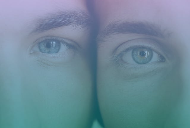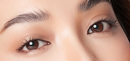
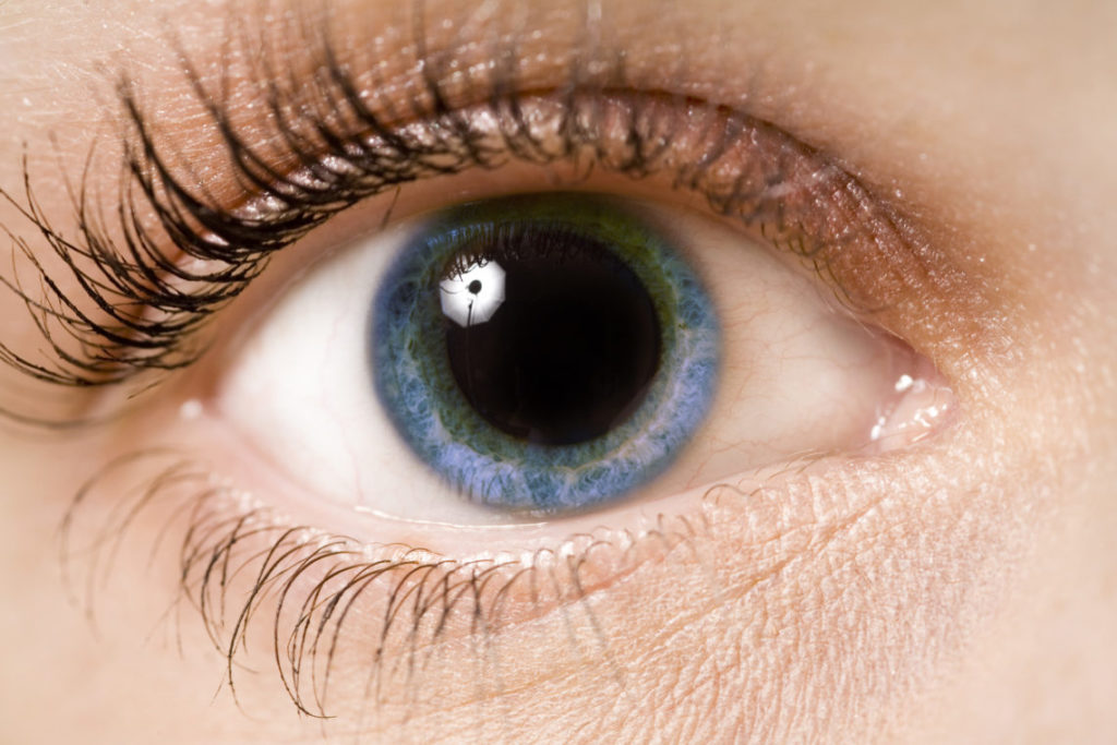
There’s an age-old saying that the eyes are the windows to a person’s soul. While it may not be a literal window into the soul itself, the pupil (the opening in the centre of your eye) does allow an eye doctor a glimpse into the internal structures of your body. So how come your doctor always wants to dilate your eyes?
Open Wide!
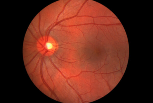
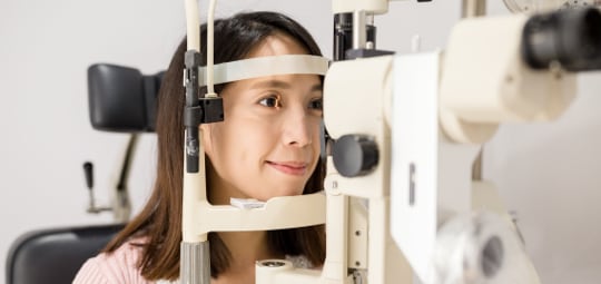
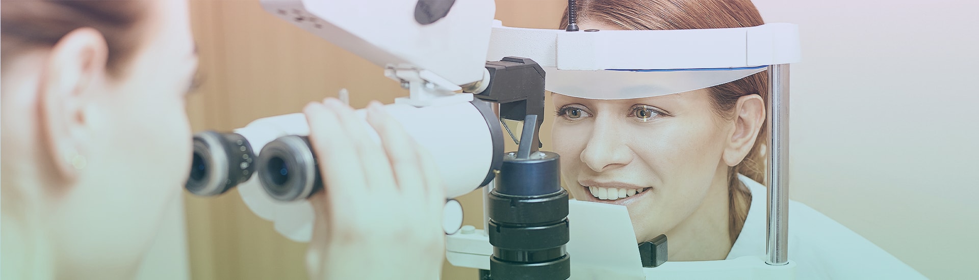
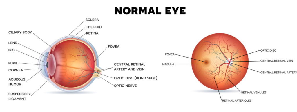
When dilated, the pupil widens and allows your eye doctor to see more than is possible in a normal eye exam, like a better view of the optic nerve, retina, arteries, veins and the macula.
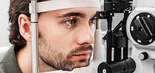
A dilated eye exam allows your eye doctor to diagnose common systemic conditions and eye diseases. Many systemic conditions such as diabetes, high blood pressure and elevated intracranial pressure are often first diagnosed at an eye exam because of signs visible inside the eye. This is why eye doctors often want to dilate your eyes so that they can make sure there is no early evidence of these conditions.
As annoying as the effects of dilated eyes can be, this only lasts a few hours and is worth the results that a truly comprehensive eye exam can provide.
What are the Benefits of a Retinal Photo?
Just like any photograph, a retinal photo captures a moment in time. The reason this is so helpful is because a photo can be used as a point of comparison for future examinations. Photos can be used in a side-by-side comparison every year to detect and monitor subtle (early) changes that may be occurring in your eyes.
Retinal photographs are also an invaluable educational tool that can help your eye doctor to better explain the state of your health and wellbeing. By reviewing your images with your doctor, they can point out changes or areas to observe and also explain treatment options. Another age-old saying is that knowledge is power, so the more educated you are about eye health, the better you’ll understand what is being recommended to you and why it’s important.